Orthopaedic doctors are medical professionals who specialize in the diagnosis, treatment, and prevention of musculoskeletal disorders and injuries. They are trained in both surgical and non-surgical techniques to treat conditions affecting bones, joints, muscles, ligaments, and tendons. The best orthopaedic doctors in chennai may also work in sports medicine and rehabilitation, providing comprehensive care to individuals suffering from musculoskeletal issues. Whether dealing with complex surgeries or conservative treatments, the best ortho specialist in chennai ensures optimal care and recovery for their patients.
Understanding the Role of an Orthopaedic Surgeon
Orthopaedic surgeons are medical professionals who specialize in diagnosing, treating, and managing conditions related to the musculoskeletal system. This system includes bones, joints, ligaments, tendons, and muscles. Their role involves:
- Diagnosis: Identifying musculoskeletal issues through physical examinations, imaging tests (like X-rays or MRIs), and patient history.
- Treatment: Providing both surgical and non-surgical treatments, including medication, physical therapy, and lifestyle modifications.
- Surgery: Performing operations to correct problems such as fractures, joint issues, or deformities. Common procedures include joint replacements, arthroscopy, and spinal surgery.
- Rehabilitation: Assisting in the recovery process, ensuring proper healing and functional restoration through rehabilitation programs and follow-up care.
- Prevention: Advising patients on injury prevention, exercise, and ergonomics to maintain musculoskeletal health.
Best ortho surgeon in chennai play a crucial role in helping patients regain mobility and improve their quality of life.
10 Best Orthopaedic Doctors in Chennai
There are some credible Orthopaedic Doctors who provide comprehensive treatments with cutting-edge techniques. Some of them include:
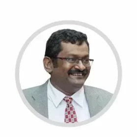
Dr. L. Bharath
Dr. L. Bharath is listed among the best orthopaedic doctors in chennai. He specializes in Knee & Hip surgery including primary, complex & revision (previously failed) joint replacements, sports injuries & arthroscopic (keyhole) procedures. He has performed 10000 surgeries and stands out as among the best Orthopaedic Surgeons in India with his extensive focus on upgrading himself on procedures and operations, in particular, Joint replacement surgery.
- Years of Experience: Over 20+ years
- Qualifications: MBBS, MS (Orthopaedics), DNB (Orthopaedics)
- Available Time: Mon-Sat 9 am to 8 pm | Sun – 10 am to 3 pm
- Address: Flat-A Ground Floor, Balaji Villa,New Door No.38/1, Old Door No.9/1, Rajaratnam Street, Kilpauk, Chennai-600010

Prof. Dr. S. Sundar
Prof. Dr. S. Sundar at VS Group of Hospitals is the Medical Director here. He is popularly known as the best Knee Replacement Surgeon in Chennai and has an extended year of experience with a specialization in knee, shoulder, and hip surgery in Chennai. He had dedicated himself to using state-of-the-art treatments using emphasized modalities to provide optimal results in joint replacements, fracture fixation, and arthroscopic surgeries for sports injuries, etc.
- Years of Experience: Over 30+ years
- Qualifications: MBBS, MS (Orthopaedics), MCh (Orthopaedics)
- Available Time: 24X7
- Address: 815/306, Poonamalle High Rd, Kilpauk, Chennai – 600010.
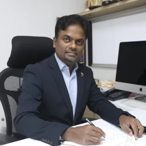
Dr. Prakash Selvam
Dr. Prakash Selvam is the Chairman and Managing Director of CTS Specialty Hospital in Chennai, India. He is one of the best orthopaedic doctors in Chennai with expertise in trauma, spine surgery, arthroscopy, joint replacement, and sports medicine. Dr. Selvam’s innovative surgical approach and extensive experience have made him a prominent figure in orthopedic medicine, contributing significantly to its advancement.
- Years of Experience: Over 15+ years
- Qualifications: MBBS, MS (Orthopaedics), DNB (Orthopaedics)
- Available Time: 24×7
- Address: V2, Plot 4047, 4th Main Rd, V Block, Anna Nagar, Chennai, Tamil Nadu 600040.
Dr. Arun Kumar
Dr. Arun Kumar stands out among the best orthopaedic doctors in chennai for his impressive qualifications, including FRCS in Trauma & Orthopaedics from the prestigious Royal College of Surgeons, Edinburgh. Serving at Medway Hospital, Dr. Kumar is known for his expertise in trauma surgery, joint replacement, and complex musculoskeletal disorders. His international training in Liverpool and extensive experience have made him a highly respected best ortho surgeon in Chennai. As a compassionate orthopedic specialist in Chennai, Dr. Arun Kumar tailors his treatments to meet the individual needs of each patient, ensuring optimal outcomes and faster recovery.
- Years of Experience: Over 22+ years
- Qualifications: MBBS, Diploma – Orthopedics, FRCS – Trauma & Orthopaedics
- Available Time: 24/7
- Address: No.Pc-7, 4th Block, Nolambur, Bharathi Salai, Mogappair West, Chennai, Tamil Nadu 600037

Dr. Karthik P Reddy
Dr. Karthik P Reddy is regarded as one of the best ortho specialists in Chennai, with a focus on knee replacements and advanced orthopaedic treatments. He practices at the renowned Knee Replacement Hospital, where his in-depth understanding of knee disorders has earned him a stellar reputation. Whether performing minimally invasive knee surgeries or treating complex cases, Dr. Reddy’s commitment to excellence ensures that patients receive top-notch care. As one of the best orthopedic surgeon in Chennai, Dr. Reddy is known for his precision, cutting-edge techniques, and personalized approach, making him a trusted name among patients seeking expert treatment.
- Years of Experience: Over 10+ years
- Qualifications: MBBS, MS Orthopaedics
- Available Time: 24X7
- Address: No.37/39, AH block, 4th Avenue, Shanthi colony, Anna nagar, Chennai- 600040

Dr. Madan Mohan Reddy
At Sunway Medical Center, Dr. Madan Mohan Reddy is highly regarded for his expertise in minimally invasive orthopaedic procedures. As one of the best orthopaedic doctors in Chennai, he is known for his skill in performing surgeries with precision, ensuring faster recovery times and minimal scarring.
- Years of Experience: Over 20+ years
- Qualifications: MBBS, MS (Orthopaedics), DNB (Orthopaedics), Fellowship in Joint Replacement
- Available Time: Mon – Sat : 11:00 AM – 7:00 PM (Sunday Emergency only)
- Address: No.37/39, AH block, 4th Avenue, Shanthi colony, Anna nagar, Chennai- 600040
Dr. Omer Sheriff
Dr. Omer Sheriff is recognized as one of the best orthopaedic doctors in chennai, specializing in joint replacement and complex orthopaedic procedures. With a distinguished academic background and a Fellowship in Joint Replacement, Dr. Sheriff has garnered a reputation for delivering exceptional care at Meridian Hospital. His expertise spans knee and hip replacement surgeries, as well as sports injuries and trauma management. As a best orthopedic surgeon in Chennai, Dr. Sheriff emphasizes personalized treatment plans and strives to achieve the highest standards of care for his patients. His advanced techniques and compassionate approach make him a trusted orthopedic specialist in Chennai.
- Years of Experience: over 24+ years
- Qualifications: MBBS, D.Ortho, M.S. Ortho, Fellowship in Joint Replacement
- Available Time: 24X7
- Address: New No. 85, Royapettah High Road, Royapettah, Chennai – 600014

Dr. Subramaniam S
Dr. Subramaniam S is an Orthopaedic Surgeon at Medway Hospitals, with a strong background in Orthopaedics. He practices at the Kodambakkam branch in Chennai, where he provides expert care in managing a variety of Orthopaedic Conditions .He holds a solid educational foundation, having completed his MBBS and D.Ortho. Additionally, he has earned a Fellowship in the Association for the Study of Internal Fixation (FASIF) from the UK, which further enhances his expertise in orthopaedic surgery. He is dedicated to offering High-Quality Patient Care, focusing on the Latest Techniques in Orthopaedic Surgery.
- Qualification: MBBS, Dip (Ortho), FASIF (UK)
- Speciality: Orthopaedics
- Location: Kodambakkam
Dr. Vigneshwaran P
Dr. Vigneshwaran P is a dedicated Consultant Orthopaedic Surgeon at Medway Hospitals, Kodambakkam, Chennai. He specializes in Orthopaedics, providing expert care in treating a wide range of Musculoskeletal conditions. He holds a solid educational background, with an MBBS and MS in Orthopaedics. His strong commitment to excellence in Orthopaedic Care ensures that his patients receive the highest quality treatment. He focuses on delivering patient-centered care, utilizing the latest advancements in Orthopaedic Surgery to achieve the best outcomes for his patients.
- Qualification : MBBS., MS (Ortho)
- Speciality : Orthopaedics
- Location : Kodambakkam
Why Chennai is a Top Choice for Orthopedic Care
- Highly Qualified Specialists: Chennai boasts renowned orthopedic surgeons with advanced training and extensive experience.
- State-of-the-Art Facilities: Leading hospitals are equipped with the latest technology and surgical techniques.
- Affordable Care: High-quality treatment at competitive costs compared to international standards.
- Comprehensive Services: Wide range of orthopedic procedures, from joint replacements to sports medicine.
- Accredited Hospitals: Many hospitals have national and international accreditations, ensuring high standards of care.
- Reputation for Excellence: Chennai is recognized for its successful outcomes and patient satisfaction in orthopedic treatments.

Conditions
- Arthritis and Joint Pain: One of the most common conditions treated by the Best Orthopaedic Doctors in Chennai, arthritis affects the joints, causing stiffness, swelling, and pain. These specialists provide long-term management plans that include medication, physical therapy, and in some cases, joint replacement surgeries.
- Fractures and Dislocations: The best ortho doctor Chennai handles everything from simple fractures to complex bone injuries using advanced imaging and surgical techniques. Quick and accurate treatment helps prevent long-term damage and ensures proper healing.
- Spine Disorders: Conditions like herniated discs, scoliosis, and spinal stenosis are managed through both conservative and surgical methods. The Best Orthopaedic Doctors in Chennai focus on pain relief, mobility restoration, and spinal stability using the latest tools and procedures.
- Sports Injuries: The best ortho doctor Chennai is equipped to manage ligament tears, tendon injuries, and stress fractures common among athletes. Their goal is to ensure safe, swift recovery while minimizing the risk of future injuries.
Conditions Treated by Orthopedic Surgeon
- Joint Disorders: The best orthopaedic doctors in chennai specialize in treating arthritis, osteoarthritis, and rheumatoid arthritis, offering both surgical and non-surgical solutions.
- Sports Injuries: From ACL tears to rotator cuff injuries, best orthopedic surgeons in Chennai are skilled in treating a wide range of sports-related injuries.
- Spinal Conditions: Leading surgeons in Chennai provide advanced treatments for herniated discs, scoliosis, and spinal stenosis.
- Fractures and Trauma: The best orthopaedic doctors in chennai offer expert care for fractures, dislocations, and other traumatic injuries.
- Pediatric Orthopedics: Top specialists in Chennai also treat congenital and developmental orthopedic issues in children, ensuring comprehensive care from infancy to adolescence.
Diseases
- Osteoporosis: Treated extensively by the Best Orthopaedic Doctors in Chennai, osteoporosis is a condition where bones become brittle and fragile due to loss of tissue. These doctors use bone density tests and personalized care plans, including medications and lifestyle changes, to reduce fracture risk.
- Osteoarthritis: This degenerative joint disease causes pain, stiffness, and reduced movement, especially in the knees, hips, and hands. The best ortho doctor chennai provides targeted therapies such as viscosupplementation, physiotherapy, or joint replacement surgery based on severity.
- Rheumatoid Arthritis: Managed by the Best Orthopaedic Doctors in Chennai, this autoimmune disease leads to joint inflammation and deformity if left untreated. Treatment involves immunosuppressants, joint protection strategies, and timely surgical intervention when needed.
- Bone Tumors: Whether benign or malignant, bone tumors require specialized care from the best ortho doctor chennai who collaborates with oncologists for surgical removal and post-treatment rehabilitation. Early diagnosis ensures better outcomes and reduced complications.
- Infectious Arthritis: The Best Orthopaedic Doctors in Chennai address joint infections caused by bacteria, viruses, or fungi with prompt diagnosis, antibiotics, and drainage procedures to prevent joint destruction.
- Avascular Necrosis: Commonly treated by the best ortho doctor chennai, this condition occurs when blood supply to the bone is disrupted, often affecting the hip. Advanced imaging and surgical options like core decompression or joint replacement are used for effective management.
Treatments
- Joint Replacement Surgery: Performed by the Best Orthopaedic Doctors in Chennai, joint replacement is recommended when conservative treatments fail to relieve severe arthritis pain. Advanced implants and minimally invasive techniques are used to restore mobility and improve quality of life.
- Arthroscopy: The best orthopedic doctor in chennai uses arthroscopy to diagnose and treat joint problems through small incisions with minimal tissue damage. This technique is effective for meniscus tears, ligament injuries, and cartilage repair, enabling faster recovery.
- Fracture Management: The Best Orthopaedic Doctors in Chennai provide personalized treatment plans using modern fixation devices like plates, screws, and rods. Their goal is to ensure proper bone alignment and speedy healing, reducing long-term complications.
- Spine Surgery: The best orthopedic doctor in chennai handles conditions such as slipped discs, spinal stenosis, and deformities with precision-guided surgical methods. These procedures are often assisted by robotics or navigation systems to enhance accuracy and patient outcomes.
- Sports Injury Rehabilitation: The Best Orthopaedic Doctors in Chennai combine physical therapy, strength training, and surgical repair when needed. Their holistic approach helps athletes return to their peak performance while minimizing re-injury risk.
Conclusion
Dr. L. Bharath is widely regarded as one of the best orthopaedic doctors in Chennai, particularly for his expertise in knee replacement surgery. With over 8000 successful surgeries and a focus on fast-track recovery protocols, Dr. Bharath combines advanced techniques with a compassionate approach. His personalized treatment plans and emphasis on post-operative rehabilitation make him a top choice for patients. As a best ortho specialist in Chennai, Dr. Bharath’s association with Bharath Orthopaedics Hospital further solidifies his reputation as a trusted and skilled professional in the field of orthopaedic care.
Read also: Hip Orthopedic Doctor In Chennai
